
- How Is Lumbar Spondylolisthesis Diagnosed?
- What Is Evaluated During a Lumbar Spondylolisthesis Examination?
- Can Lumbar Spondylolisthesis Be Seen on an X-Ray?
- Can Lumbar Spondylolisthesis Be Detected with MRI?
- What Does Computed Tomography Show in Lumbar Spondylolisthesis?
- How Is the Grade of Lumbar Spondylolisthesis Determined?
- How Is Spondylolisthesis Diagnosed?
- How Is Nerve Compression Evaluated in Lumbar Spondylolisthesis?
- How Is Lumbar Spondylolisthesis Distinguished from a Herniated Disc?
- Physical and Neurological Examination in the Diagnosis of Lumbar Spondylolisthesis
- Why Might Standing X-Rays Be Requested in Lumbar Spondylolisthesis?
- What Is the Difference Between MRI and X-Ray in Lumbar Spondylolisthesis?
- How Is Treatment Planned After a Diagnosis of Lumbar Spondylolisthesis?
- How Is the Progression of Lumbar Spondylolisthesis Monitored?
- Frequently Asked Questions About the Diagnosis of Lumbar Spondylolisthesis
How Is Lumbar Spondylolisthesis Diagnosed?
The first stage in evaluating how lumbar spondylolisthesis is diagnosed is taking a detailed medical history. The duration of lower back pain, whether the pain increases with standing or walking, whether it radiates to the hip or leg, and whether neurological complaints such as numbness are present are investigated. In some people, the slippage may remain asymptomatic for a long time, while in others even a low-grade slippage may cause complaints that affect daily life. Therefore, evaluating the condition solely by looking at the amount of slippage on imaging is not sufficient.
After a detailed lumbar spondylolisthesis examination, imaging methods are used when necessary. Standing X-rays are particularly important because they show the alignment of the vertebrae under load. When more information is needed about nerve tissues and intervertebral discs, magnetic resonance imaging may be added to the lumbar spondylolisthesis imaging process. Computed tomography may be used when a detailed assessment of bone structures is required.
When the slippage occurs in the lumbar vertebrae, the condition is referred to as lumbar spondylolisthesis. During diagnosis, not only the displacement of one vertebra relative to another is assessed, but also the level at which it occurs, whether there is instability that increases with movement, and whether the neural canals are affected. This allows imaging results to be interpreted together with the patient's examination findings.
What Is Evaluated During a Lumbar Spondylolisthesis Examination?
During the examination, the patient's posture, the natural curvature of the lower back, walking pattern, and spinal movements are evaluated. It is assessed whether the pain changes when the lower back is moved forward, backward, and sideways. Complaints related to lumbar spondylolisthesis may become more noticeable especially during prolonged standing, backward bending, or long-distance walking; however, these findings are not the same in every patient.
During a lumbar spondylolisthesis examination, the strength of the leg muscles, sensory function, and reflexes are also checked. If the nerve roots are affected, numbness, tingling, weakness, or pain may occur in the leg. The doctor may also ask the person to walk on their heels and toes to evaluate the function of specific nerve roots.

No single lumbar spondylolisthesis test can establish a definitive diagnosis on its own. Findings obtained during the physical examination should be evaluated together with X-ray, magnetic resonance imaging, or, when necessary, computed tomography findings. This approach helps determine whether the pain is truly caused by vertebral slippage or by another spinal problem.
Can Lumbar Spondylolisthesis Be Seen on an X-Ray?
Lumbar spondylolisthesis X-ray imaging can often show the slippage directly. In particular, a lateral lumbar X-ray is one of the basic imaging methods used to assess how far one vertebra has displaced relative to the vertebra immediately below it. X-rays can also provide information about the general alignment of the vertebrae, narrowing of disc spaces, degenerative changes, and spinal posture.
Images taken while standing are important because they show the actual mechanical condition of the spine under body weight. Slippage that may not be clearly visible on some images taken while lying down can be easier to detect under load. When necessary, flexion and extension X-rays taken while bending forward and backward may also help assess whether there is greater-than-normal movement between the vertebrae.
Spondylolisthesis X-ray evaluation is not performed only to demonstrate the presence of slippage. The percentage of slippage can be calculated, overall spinal balance can be examined, and comparisons can be made with previous images during follow-up. However, because X-rays do not provide detailed information about nerves, intervertebral discs, and other soft tissues, additional imaging may be required in some patients.
Can Lumbar Spondylolisthesis Be Detected with MRI?
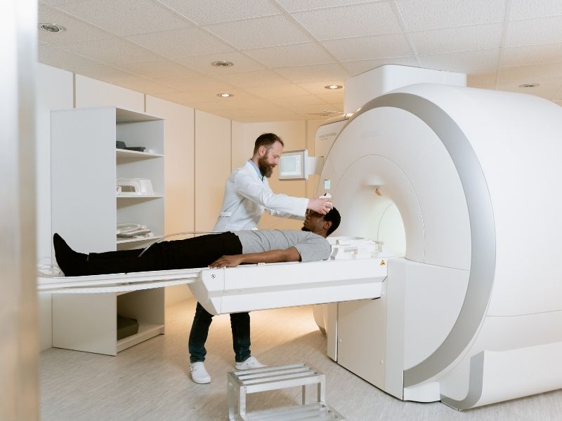
Lumbar spondylolisthesis MRI can show the slippage, but one of the most important advantages of magnetic resonance imaging is its ability to display nerve tissues and the soft structures of the spine in detail. The condition of the discs between the vertebrae, narrowing of the neural canals, compression of the nerve roots, and accompanying problems such as a herniated disc can be evaluated on MRI images.
The colloquial term lumbar spondylolisthesis MRI scan refers to the same examination. MRI becomes particularly important in patients with pain radiating to the leg, numbness, tingling, weakness in the legs while walking, or neurological loss. This is because the examination allows a more detailed assessment of whether the patient's complaints are related to nerve compression caused by the slippage rather than the vertebral displacement itself.
Spondylolisthesis MRI results should always be interpreted together with examination and X-ray findings. Because MRI is generally performed in the lying position, mobility that appears or increases under load may not always be seen to the same degree. Therefore, in some patients, standing X-rays and MRI are used as complementary examinations.
What Does Computed Tomography Show in Lumbar Spondylolisthesis?
Lumbar spondylolisthesis CT provides highly detailed imaging of bone structures. Computed tomography can provide important information especially when a crack or bone defect is suspected in the pars interarticularis, a region located at the back of the vertebra. This is particularly important in the evaluation of certain types of spondylolisthesis that develop at a young age.
On lumbar spondylolisthesis CT scans, the bony anatomy of the vertebrae, joint surfaces, bone spurs, and structural changes accompanying the slippage can be examined in detail. In some patients who require surgical planning, CT may also be used to better understand the three-dimensional anatomy of the bone structures.
Computed tomography is not routinely required for every patient. MRI provides more detailed information about the nerve roots and discs, whereas CT is stronger for assessing bone tissue. Therefore, the imaging method to be used is determined according to the patient's age, complaints, examination findings, and the results of previous imaging studies.
How Is the Grade of Lumbar Spondylolisthesis Determined?
Grades of lumbar spondylolisthesis are determined according to how far the upper vertebra has moved forward over the vertebra below it. In one of the most commonly used classifications, slippage of up to 25% is considered grade 1, 25% to 50% grade 2, 50% to 75% grade 3, and 75% to 100% grade 4. Advanced cases in which the vertebra has completely displaced forward may be defined as spondyloptosis.
The degree of slippage is usually calculated on a lateral X-ray. However, it should not be assumed that a high-grade slippage will necessarily cause very severe pain or that a low-grade slippage is insignificant. The patient's symptoms, nerve compression, the behavior of the spine during movement, and the effects on daily life should be evaluated together.
During follow-up, comparing previous and new X-rays can show whether the slippage has progressed. Radiological follow-up may become particularly important in growing patients, individuals with marked instability, or patients whose complaints increase over time.
How Is Spondylolisthesis Diagnosed?
Diagnosis of spondylolisthesis is made by demonstrating that one vertebra has displaced relative to the adjacent vertebra and evaluating the clinical significance of this condition. During the diagnostic process, the patient's age, the onset of complaints, level of physical activity, previous trauma, and degenerative changes in the spine are taken into account. This is because spondylolisthesis is a spinal problem that can occur for different reasons.
For a spondylolisthesis diagnosis, most patients undergo X-ray evaluation after a physical and neurological examination. If nerve compression is suspected or if a detailed evaluation of the discs and spinal canal is required, MRI may be requested. Computed tomography may be used when more detailed information about bone structures is needed.
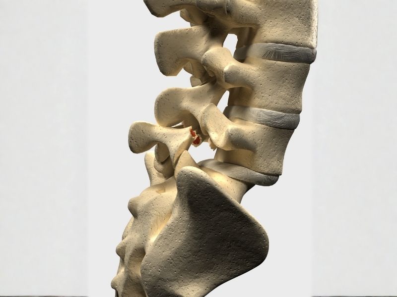
During diagnosis, particular attention is paid to whether the slippage seen on imaging is related to the patient's complaints. This is because in some people, spondylolisthesis may be detected incidentally on imaging performed for another reason, and the person may experience no symptoms. Therefore, the imaging result alone does not determine the treatment decision.
How Is Nerve Compression Evaluated in Lumbar Spondylolisthesis?
Nerve compression in lumbar spondylolisthesis may develop due to narrowing of the spaces through which the nerve roots pass as a result of vertebral displacement or accompanying degenerative changes. Patients may experience pain radiating from the lower back to the hip and leg, numbness, tingling, a burning sensation, or reduced strength in certain muscle groups.
During the neurological examination, muscle strength, sensation, and reflexes are evaluated in detail. The possible level of nerve root compression is assessed together with the distribution of pain and numbness in the leg. A reduced walking distance or the development of pain and weakness in the legs while walking may suggest narrowing of the spinal canal.
MRI has an important role in imaging nerve compression. The width of the canals through which the nerve roots pass, the condition of the discs, and other structural changes accompanying the slippage can be seen in the same examination. New-onset problems with bladder or bowel control, numbness around the groin, or rapidly progressive significant weakness are findings that require prompt medical evaluation.
How Is Lumbar Spondylolisthesis Distinguished from a Herniated Disc?
Because lumbar spondylolisthesis and a herniated disc can cause similar complaints, a definitive distinction cannot be made based only on the location of the pain. In both conditions, lower back pain, pain radiating to the hip or leg, numbness, and tingling may occur. In addition, some patients may have both conditions at the same time.
In a herniated disc, the disc between the vertebrae protrudes toward the neural tissue, whereas in lumbar spondylolisthesis the normal alignment between the vertebrae is disrupted. In evaluating how lumbar spondylolisthesis is identified, X-rays show vertebral alignment while MRI provides detailed information about the discs and neural structures. Therefore, examining physical examination findings together with imaging findings is important in distinguishing between the two conditions.
After a definitive lumbar spondylolisthesis diagnosis is made, an attempt is made to determine whether the patient's complaints are due to the slippage, accompanying spinal canal narrowing, a herniated disc, or a combination of several of these problems. The source of pain should be identified as clearly as possible so that the treatment plan can be created appropriately.
Physical and Neurological Examination in the Diagnosis of Lumbar Spondylolisthesis
During the physical examination, the shape of the spine, lower back movements, muscle tenderness, and the person's walking pattern are evaluated. In some patients, the natural inward curve of the lower back may increase, or a step-off that can be felt by hand may be noticed at a certain level of the spine. However, the absence of these findings does not rule out lumbar spondylolisthesis.
During the neurological assessment, leg muscle strength, reflexes, and sensation are checked. Movements such as lifting the ankle upward, moving the big toe, or rising onto the toes can provide information about the functions of different nerve roots. Even if the patient's complaints consist only of pain, neurological assessment remains an important part of the diagnosis.
No single lumbar spondylolisthesis test definitively confirms the condition. Findings obtained during the examination help determine which imaging method is necessary. Therefore, physical and neurological examinations remain fundamental stages of assessment both before imaging and while interpreting imaging results.
Why Might Standing X-Rays Be Requested in Lumbar Spondylolisthesis?
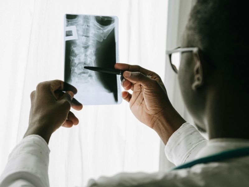
The spine is a weight-bearing structure, and some alignment problems may become more apparent when a person is standing. Therefore, performing a lumbar spondylolisthesis X-ray while standing may show the actual position of the vertebrae under body weight. A difference in the amount of slippage may be noticed especially when compared with images taken in the lying position.
The amount of slippage can be measured on a standing lateral X-ray, and the overall balance of the spine can be evaluated. If the doctor considers it necessary, flexion and extension X-rays taken while bending forward and backward may also be used. These images contribute to the assessment of whether there is instability between the vertebrae that becomes more apparent with movement.
Changes seen on standing X-rays should be evaluated together with the patient's symptoms. Detecting mobility on imaging alone does not mean that surgical treatment will be required. The treatment decision is made by considering the level of pain, neurological findings, the grade of slippage, its effects on daily life, and other imaging results together.
What Is the Difference Between MRI and X-Ray in Lumbar Spondylolisthesis?
X-ray and MRI are complementary rather than alternative methods in the evaluation of lumbar spondylolisthesis. X-rays are effective in showing vertebral alignment, the amount of slippage, and mechanical changes that occur under load. Standing lateral images are particularly useful for grading the slippage.
The main advantage of lumbar spondylolisthesis MRI is that it provides detailed information about the discs, nerve roots, spinal canal, and other soft tissues. In patients with pain radiating to the leg, numbness, tingling, or weakness, MRI may be important for evaluating the condition of the neural tissues.
Therefore, the lumbar spondylolisthesis imaging plan may vary from patient to patient. In some people, X-rays provide sufficient basic information for diagnosis, while MRI may be required in patients with suspected nerve compression or an additional spinal problem. CT may also be added to the evaluation when a detailed examination of the bone anatomy is needed.
How Is Treatment Planned After a Diagnosis of Lumbar Spondylolisthesis?
After the diagnosis of lumbar spondylolisthesis is confirmed, treatment is not determined solely according to the degree of slippage seen on imaging. The patient's pain level, limitations in daily activities, neurological findings, age, spinal stability, and the course of the slippage over time are evaluated together. Therefore, different treatment options may be considered for two people with the same degree of slippage.
In some patients with mild or moderate complaints and no progressive neurological loss, nonsurgical methods may be applied first. Activity modification, medications considered appropriate by the doctor, and individualized exercise or physical therapy programs may be part of this approach. The aim is to control pain and preserve the person's daily functional abilities.
If there is progressive weakness, significant nerve compression, severe pain that persists despite treatment, or clinically significant spinal instability, surgical options may be considered. Whether surgery is necessary and which technique should be used must be evaluated separately for each patient. An imaging finding alone does not constitute a decision for surgery.

How Is the Progression of Lumbar Spondylolisthesis Monitored?
In monitoring lumbar spondylolisthesis, changes in the patient's complaints are as important as imaging results. Comparing new X-rays with previous ones can show whether the amount of slippage has increased. Follow-up intervals are planned individually, especially in young patients who are still growing or in people with suspected mechanical instability.
Grades of lumbar spondylolisthesis may be remeasured during follow-up, but focusing only on radiological change is not sufficient. Symptoms such as increased pain, reduced walking distance, newly developed numbness, or loss of muscle strength are also important in evaluating the clinical course of the condition. If new neurological complaints develop, reevaluation may be necessary without waiting for the previously scheduled follow-up appointment.
Frequently Asked Questions About the Diagnosis of Lumbar Spondylolisthesis
Can Spondylolisthesis Occur Without Any Symptoms?
Yes, in some people, lumbar spondylolisthesis may progress without symptoms and may be detected incidentally during spondylolisthesis imaging examinations performed for another reason.
Which Doctor Should Be Consulted for Spondylolisthesis?
For spondylolisthesis diagnosis, a neurosurgery specialist can be consulted. During the specialist evaluation, a spondylolisthesis examination and necessary imaging methods are planned together.
Can Spondylolisthesis Be Diagnosed Definitively With MRI Alone?
Spondylolisthesis MRI can show the condition, but an X-ray may also be required to evaluate the degree of slippage and its status under load. In particular, spondylolisthesis MRI provides detailed information about the nerves and discs.
What Is a Dynamic X-Ray Used for in Spondylolisthesis?
Spondylolisthesis X-ray images taken while bending forward and backward help evaluate whether there is increased slippage or instability between the vertebrae with movement. In some patients, this examination may provide additional information for the diagnosis of vertebral slippage.
Is Computed Tomography Necessary for Every Patient With Spondylolisthesis?
No, spondylolisthesis computed tomography is not necessary for every patient. Spondylolisthesis CT may be preferred especially for evaluating bone structures, the pars region, and anatomy before surgery.
Can the Degree of Spondylolisthesis Change Over Time?
In some patients, degrees of spondylolisthesis may progress over time and can be monitored with follow-up X-rays. Re-evaluation is especially important if new pain, numbness, or loss of strength develops.
7. What Findings Require Emergency Evaluation in Spondylolisthesis?
Progressive muscle weakness, newly developed problems with bladder or bowel control, and numbness around the groin may require emergency evaluation for nerve compression in spondylolisthesis. If these symptoms occur, medical evaluation should be performed without delay.