What Is Spinal Stenosis?
The spine is a very important structure that supports the body and enables it to remain upright; it is especially essential for our movements.
The spine also contains the canal located in the center of the vertebrae, through which the spinal cord passes, known as the spinal canal. The spinal cord in the lower back carries the nerves that provide sensation and strength to the legs. When the canal between the vertebrae narrows, the spinal cord and nerve roots passing through it become compressed. This is the most common cause of spinal stenosis.
As age advances, the discs between the vertebrae begin to lose water, causing the space between the vertebrae to narrow. In response to this condition, extra bone formation, in other words calcification, is observed in the spine and the small joints of the lower back. In addition, the yellow ligaments connecting the vertebrae tend to thicken toward the canal, and the disc protrudes into the canal. As a result of all these symptoms, the spinal canal narrows to a certain extent. In other words, spinal canal stenosis is the narrowing over time of the thin canal located in the middle of the spinal bones, through which the spinal cord and nerves pass, resulting in pressure on the spinal cord and nerves. Because of this pressure, the spinal cord and nerves become unable to perform their normal functions.
Various diagnostic methods are used to distinguish spinal stenosis from other diseases. The first step in diagnosing spinal stenosis is obtaining information about the patient's history and complaints. After collecting data such as when the complaints began, where the pain is felt, and other health problems, the patient is examined in the second stage. During the physical examination, a detailed neurological examination is performed by evaluating the patient's strength, sensation, and reflexes. As a third step, imaging tests are requested from the patient. X-ray is one of the most basic diagnostic tools for spinal stenosis. The stenosis can be clearly detected with radiographs taken using X-rays. In addition; the canal diameter can be measured with MRI or tomography imaging in spinal stenosis, and the diagnosis can be made easily.
Narrowing of the lumbar canal especially occurs after the age of 50 due to the discs losing their elasticity. Although rare, it can also be seen in younger people with a congenital predisposition. It is more common in men than in women. Some patients with spinal stenosis in the lumbar spine may also have stenosis in the cervical spine.
Symptoms of Spinal Stenosis
Narrowing of the canal does not always cause symptoms. Many cases have been observed in which spinal stenosis is significant but does not cause any complaints in the person.
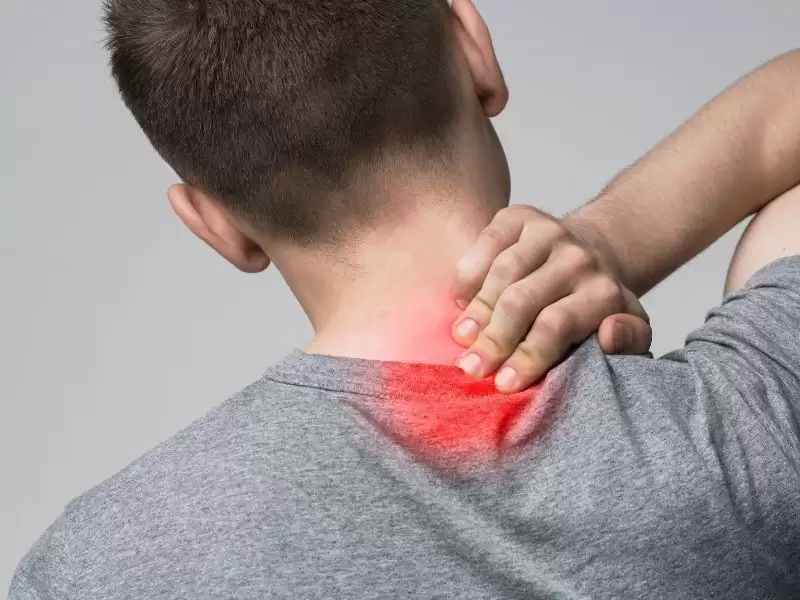
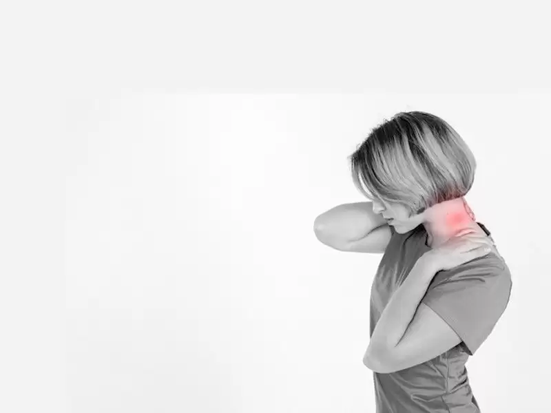
Symptoms of spinal stenosis are often confused with a herniated disc. However; the most common symptom of spinal stenosis is that all symptoms progressively affect both legs and cause complaints. Lower back pain, pain in both legs, numbness in both legs, tingling, swelling, and edema are the main symptoms of spinal stenosis. The fact that these complaints progressively increase in both legs distinguishes spinal stenosis from a herniated disc. In a herniated disc, only a single nerve on the right or left side is compressed.
Patients with complaints of spinal stenosis feel pain and numbness in the legs and hips even if they walk a short distance. These pains are accompanied by neurogenic claudication, that is, weakness in the muscles.
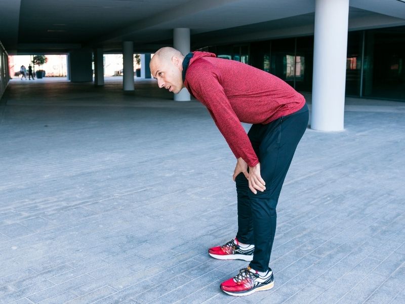
A patient experiencing muscle weakness feels the need to sit down immediately even after walking a short distance; otherwise, they feel as if they are going to fall. These types of complaints increase even more when standing for a long time. In positions such as bending forward, sitting, and lying down, however, the canal widens and a reduction in complaints is observed. When a patient with spinal canal narrowing is asked to walk, their pain begins to intensify. The patient tends to walk leaning forward. The reason for this is that the canal widens in the bent position and the pain decreases. This is a typical condition seen in patients with spinal stenosis and is the most important and distinctive symptom.
The patient's walking distance gradually begins to decrease due to spinal stenosis. Spinal stenosis limits the person's movements and causes disruption in daily life. The most troublesome aspect of spinal stenosis is the progressive worsening of the symptoms. The distance the patient can walk without stopping becomes shorter day by day. In advanced cases, the problems make themselves felt even at home in the form of numbness in the legs.
Although it is a rare symptom, if spinal stenosis progresses significantly and reaches the final stages, weakness severe enough to cause paralysis in the muscles may be observed. It may even progress to complaints such as loss of bowel control and urinary incontinence due to muscle weakness. In such a case, surgical intervention should be sought without delay.
Symptoms of spinal stenosis generally respond to conservative treatment. Surgical intervention is sought for lower back pain, leg pain, and neurogenic claudication (muscle weakness) that cannot be treated with physical therapy, lumbar corsets, and back exercises. For patients who do not respond to conservative treatments, the most definitive treatment is spinal stenosis surgery. In spinal stenosis surgery, the spinal cord and nerve roots are relieved of pressure and the spinal alignment is restored. By widening the canal and thus eliminating the pressure caused by the stenosis, the patient's complaints also disappear. In the long-term results of spinal stenosis surgery, an increase in patients' walking duration is observed and they are able to move much more comfortably compared to before the surgery.