Spinal cord tumors are masses arising from the spinal cord tissue and surrounding structures and may lead to progressive neurological complaints. Findings such as back or neck pain, loss of strength in the arms and legs, numbness, or balance problems bring the diagnostic process to the agenda. At this point, the diagnosis of spinal cord tumor is made in a planned manner through detailed neurological evaluation and advanced imaging methods. Early and accurate diagnosis plays a decisive role in determining the treatment approach.
- How Is Spinal Cord Tumor Diagnosed?
- The Importance Of Physical Examination In The Diagnosis Of Spinal Cord Tumor
- Why Is MRI Necessary In The Diagnosis Of Spinal Cord Tumor?
- Which Imaging Methods Are Used In The Diagnosis Of Spinal Cord Tumor?
- Is Computed Tomography Used In The Diagnosis Of Spinal Cord Tumor?
- Is Biopsy Necessary In Spinal Cord Tumor?
- The Role Of Pathological Examination In The Diagnosis Of Spinal Cord Tumor
- How Long Does It Take To Diagnose Spinal Cord Tumor?
- How Is Differential Diagnosis Made In The Diagnosis Of Spinal Cord Tumor?
- The Importance Of Early Diagnosis In Spinal Cord Tumor
- Which Department Diagnoses Spinal Cord Tumor?
How Is Spinal Cord Tumor Diagnosed?
Diagnosis of spinal cord tumor is made by evaluating the patient’s complaints, neurological examination findings, and advanced imaging methods together. Masses arising from the spinal cord tissue and surrounding structures may produce different symptoms depending on their location and growth rate. Findings such as back and neck pain, weakness in the arms or legs, numbness, impaired walking balance, and changes in urinary control create clinical suspicion. At this point, a systematic evaluation process is initiated for the diagnosis of spinal cord tumor.
In the practice of neurosurgery, when establishing the diagnosis of spinal cord tumor, importance is given not only to imaging findings but also to the patient’s detailed history. How long the complaints have been present, whether they are progressive, and the degree of neurological loss guide the diagnostic process. The aim is to clearly reveal the tumor’s location, size, degree of pressure on the spinal cord, and its relationship with surrounding tissues. Accurate and timely diagnosis of spinal cord tumor is the main step in determining the treatment plan.
The Importance Of Physical Examination In The Diagnosis Of Spinal Cord Tumor
In the diagnosis of spinal cord tumor, physical examination constitutes the basic step of evaluation. In a detailed neurological examination, muscle strength, deep tendon reflexes, superficial and deep sensory functions, as well as balance and coordination are carefully assessed. The aim is to determine whether there is neurological loss at a certain level along the spinal cord. Findings such as one-sided loss of strength, loss of sensation at a certain sensory level, or increased reflexes may suggest spinal cord compression.
The data obtained during the examination help indicate at which segment the lesion may be located in terms of the diagnosis of spinal cord tumor. A mass located in the neck region and a mass in the back or lower back region create different clinical presentations.

For this reason, physical examination is important not only for making a preliminary diagnosis but also for determining the correct imaging area. A comprehensive neurological evaluation performed by an experienced neurosurgery specialist is an indispensable part of the diagnosis process of spinal cord tumor.
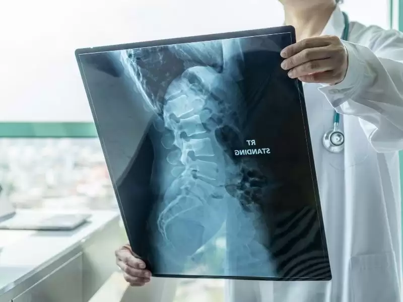
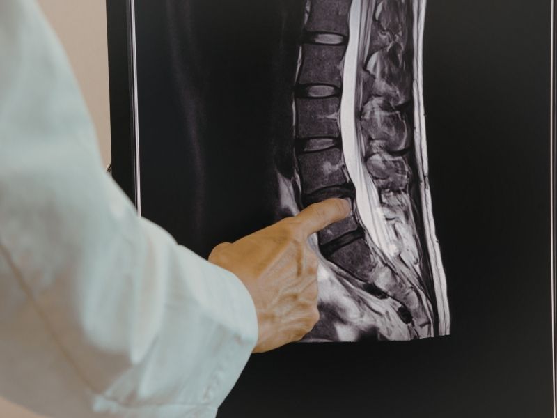
Why Is MRI Necessary In The Diagnosis Of Spinal Cord Tumor?
Magnetic resonance imaging is the gold standard method in the diagnosis process of spinal cord tumor. MRI shows the spinal cord tissue, nerve roots, and surrounding soft tissues in high resolution. In contrast-enhanced examinations, the tumor’s borders, vascularity characteristics, and relationship with the spinal cord are evaluated in detail.
The main reason why MRI is preferred for the diagnosis of spinal cord tumor is that it does not involve radiation and provides detailed multiplanar imaging. Intramedullary, intradural extramedullary, or extradural masses can be distinguished thanks to MRI. In addition, conditions such as edema, compression, and spread within the spinal cord can also be clearly seen. This information is decisive in terms of surgical planning.
Which Imaging Methods Are Used In The Diagnosis Of Spinal Cord Tumor?
Although MRI is the main method in the diagnosis process of spinal cord tumor, additional imaging techniques may be needed in some situations. Computed tomography is especially useful in evaluating bone structures. Whether there is bone erosion or structural deterioration in the spine can be examined with this method.
In some patients, whole-body scans may be performed for advanced-stage evaluation. The aim is to investigate whether there are other foci in a patient diagnosed with spinal cord tumor. Additional examinations may be planned especially when metastasis is suspected. The imaging strategy for each patient is determined individually according to the clinical picture.
Is Computed Tomography Used In The Diagnosis Of Spinal Cord Tumor?
Computed tomography is used as an auxiliary method in the diagnosis process of spinal cord tumor. Although it is not as detailed as MRI in soft tissue evaluation, it provides important information about bone structures. Collapse in the vertebral body, bone destruction, or the presence of calcification can be seen more clearly with CT.
In some patients, when MRI cannot be performed, CT myelography may be preferred as an alternative for the diagnosis of spinal cord tumor. In this method, the spinal canal is evaluated with contrast material. It may contribute to diagnosis especially in patients with implants or in those who are not suitable for MRI.
Is Biopsy Necessary In Spinal Cord Tumor?
In some situations, biopsy may be necessary while confirming the diagnosis of spinal cord tumor. Although imaging findings strongly suggest the presence of a tumor, the type of tumor can only be clarified by a tissue sample. It is especially important to determine whether the tumor is benign or malignant before surgical planning.
The decision for biopsy is made by considering the tumor’s location and the patient’s clinical condition. In some cases, the diagnosis of spinal cord tumor is confirmed directly through tissue removed during surgery. The aim is to clarify the diagnosis while minimizing the risk of damage to the spinal cord.
The Role Of Pathological Examination In The Diagnosis Of Spinal Cord Tumor
Pathological examination is the most definitive stage of the diagnosis of spinal cord tumor. The obtained tissue sample is examined under a microscope and the cellular characteristics of the tumor are evaluated. The tumor’s type, grade, and biological behavior are determined according to the pathology result.
When the diagnosis of spinal cord tumor is pathologically confirmed, the treatment plan is outlined more clearly. While surgery may be sufficient in benign tumors, additional treatment options may come to the agenda in some malignant tumors. For this reason, the pathology report is one of the main elements determining the direction of the process.
How Long Does It Take To Diagnose Spinal Cord Tumor?
The time required for the diagnosis of spinal cord tumor varies depending on when the patient presents and the existing findings. When the complaints are noticed in the early period and specialist evaluation is sought, the diagnostic process progresses more quickly. MRI and other imaging methods can usually be planned within a short time.
However, if biopsy and pathology are required before the diagnosis of spinal cord tumor is finalized, the process may take longer. On average, a preliminary diagnosis can be made within a few days after imaging, while pathological confirmation may take a few weeks. Early presentation is an important factor that shortens the diagnosis period.
How Is Differential Diagnosis Made In The Diagnosis Of Spinal Cord Tumor?
Before a diagnosis of spinal cord tumor is made, other diseases causing similar symptoms should be excluded. Spinal disc herniations, infections, demyelinating diseases, and vascular malformations may create similar neurological findings. For this reason, the differential diagnosis process is carried out carefully.
By evaluating MRI findings and the clinical picture together, the diagnosis of spinal cord tumor is clarified. Additional laboratory tests and advanced examinations may be performed when necessary. The aim is to prevent misdiagnosis and determine the most suitable treatment approach for the patient.
The Importance Of Early Diagnosis In Spinal Cord Tumor
When the diagnosis of spinal cord tumor is made in the early stage, the possibility of permanent damage caused by pressure on the spinal cord tissue decreases. The spinal cord is a sensitive neural structure, and prolonged pressure may lead to neurological problems such as permanent loss of strength, sensory deficit, and gait disturbance. For this reason, gradually increasing pain, weakness in the arms or legs, and sensory changes should be evaluated carefully.
A timely diagnosis of spinal cord tumor allows treatment planning to be carried out in a more controlled and safe manner. In situations requiring surgical intervention, procedures performed in the early stage contribute to preserving nerve tissue. In addition, early diagnosis plays a decisive role in preserving the patient’s capacity to maintain daily life activities.
Which Department Diagnoses Spinal Cord Tumor?
The diagnosis of spinal cord tumor is evaluated and confirmed by a neurosurgery specialist. The patient’s neurological examination, analysis of complaints, and planning of the necessary imaging studies are carried out within this specialty. When necessary, surgical intervention is also performed by neurosurgery. Coordinated work with radiology and pathology units is essential during the diagnostic process.
It is important for patients with suspicion of spinal cord tumor diagnosis to consult the relevant specialist without delay. A systematic evaluation process carried out by an experienced team ensures that the correct diagnosis is made and an appropriate treatment plan is established. This approach is decisive both for preserving neurological functions and for increasing treatment success.